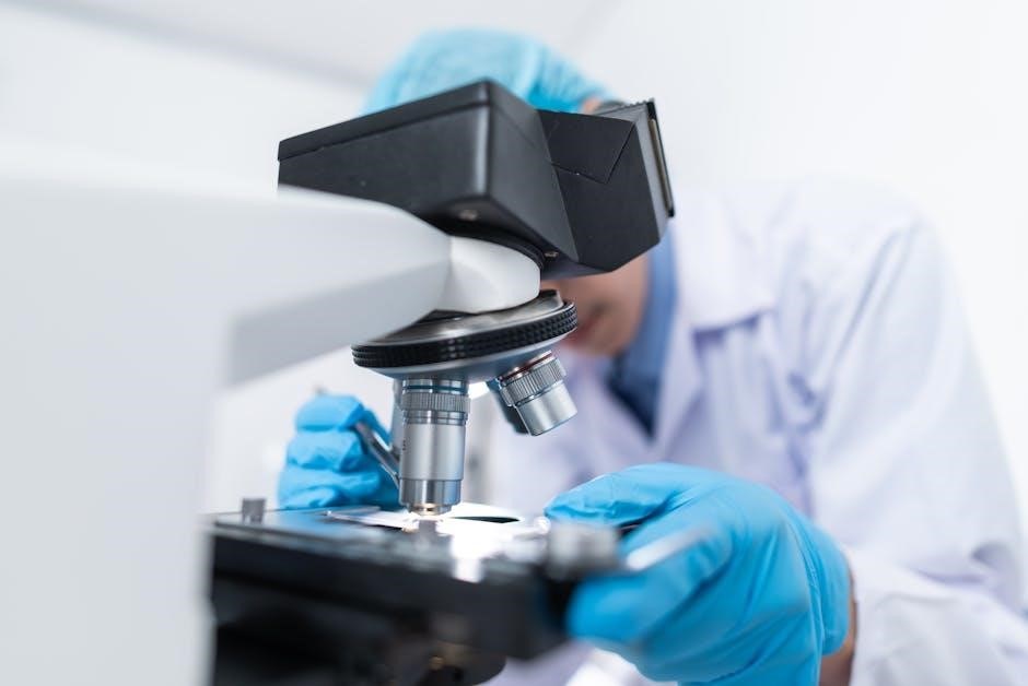
This educational resource provides a comprehensive guide to understanding microscopes‚ covering essential concepts‚ parts‚ functions‚ and calculations. Ideal for students and educators‚ it includes practical exercises and answer keys to enhance learning experiences in biology and microscopy. The PDF format ensures easy access and printing for classroom or homework use.
Why is it important to study microscope worksheets?
Studying microscope worksheets is essential for understanding the fundamental concepts of microscopy‚ enabling students to identify and describe microscope parts‚ their functions‚ and proper usage. These worksheets enhance practical skills like magnification calculations‚ specimen preparation‚ and focusing techniques. They also foster problem-solving abilities and reinforce key biological concepts‚ making them invaluable for students in science and biology. Regular practice with microscope worksheets ensures a strong foundation for future scientific studies and real-world applications in fields like medicine and research.

History of Microscopes
The microscope’s origins trace back to 1590 in the Netherlands‚ invented by Hans Jansen and his son Zacharias. Their design led to the first practical compound microscope‚ revolutionizing scientific observation.
Key inventors and their contributions to microscopy
Hans Jansen and his son Zacharias are credited with inventing the first compound microscope in 1590. Antonie van Leeuwenhoek‚ known as the “Father of Microbiology‚” improved microscope design and discovered microorganisms. Later innovators refined lenses and mechanisms‚ enhancing resolution and usability for scientific and educational purposes.
Historical milestones in microscope development
The invention of the compound microscope in 1590 revolutionized science‚ enabling detailed observations of tiny structures. In the 17th century‚ Antonie van Leeuwenhoek’s improved designs revealed microorganisms‚ advancing microbiology. The 19th century saw significant advancements with Carl Zeiss and Ernst Abbe’s lens innovations. The 20th century introduced electron microscopes‚ surpassing light microscopy limits. These milestones have transformed microscopy into an indispensable tool for scientific discovery and education‚ as highlighted in microscope worksheet resources.

How Microscopes Help Us Study Living Things
Microscopes reveal the invisible‚ allowing us to observe cells‚ microorganisms‚ and biological processes in detail‚ essential for understanding life sciences and advancing medical research and education.
Magnification and resolution: Key concepts
Magnification refers to the ability of a microscope to enlarge specimens‚ calculated by multiplying the eyepiece and objective lens magnifications. Resolution determines clarity‚ ensuring distinct images. Higher numerical aperture enhances resolution‚ allowing finer details to be observed. Proper adjustment of focus and use of immersion oil maximize these factors‚ enabling precise study of microscopic structures. Understanding these concepts is crucial for optimal microscopy results in education and research settings.
Observing cells and microorganisms
Microscopes enable detailed observation of cells and microorganisms‚ revealing structures invisible to the naked eye. Proper staining techniques enhance visibility‚ while magnification and resolution ensure clarity. Students learn to identify cell components‚ such as nuclei and mitochondria‚ and distinguish between prokaryotic and eukaryotic cells. Microorganisms like bacteria and protozoa are studied to understand their morphology and behavior. These observations are crucial for biology education‚ fostering a deeper understanding of life at the microscopic level and its significance in various scientific fields.
Labeling the Parts of a Microscope
Accurately identifying and labeling microscope components is essential for understanding their functions. Key parts include the eyepiece‚ objective lenses‚ stage‚ and focus knobs. This exercise helps students grasp microscope operation and prepare for practical experiments‚ enhancing their ability to explore microscopic structures effectively.
Identifying the main components of a compound microscope
Understanding the main parts of a compound microscope is crucial for effective use. Key components include the eyepiece‚ objective lenses‚ revolving nosepiece‚ stage‚ stage clips‚ coarse and fine focus knobs‚ arm‚ and light source. Each part plays a specific role in achieving clear magnification and proper specimen observation. Labeling these components accurately helps students develop a strong foundation in microscopy techniques and prepares them for advanced biological studies. This section provides detailed diagrams and labels for easy identification and learning.
Understanding the functions of each part
Each component of a compound microscope serves a specific function. The eyepiece magnifies the image‚ while objective lenses focus light from the specimen. The stage holds the slide‚ secured by stage clips; Coarse and fine focus knobs adjust the lens position for clarity. The light source illuminates the specimen‚ and the arm provides stability. Understanding these roles is essential for proper microscope operation and achieving accurate observations in scientific studies.
Calculating Magnification
Magnification is calculated by multiplying the eyepiece magnification by the objective lens magnification. This formula helps determine the total power of the microscope‚ essential for precise observations.
The formula for total magnification
The total magnification of a microscope is calculated by multiplying the magnification of the eyepiece by the magnification of the objective lens. This formula is expressed as:
Total Magnification = Eyepiece Magnification × Objective Lens Magnification. For example‚ if the eyepiece has a 10x magnification and the objective lens has a 40x magnification‚ the total magnification would be 400x. This calculation is essential for understanding the scale of observations made through a microscope‚ ensuring accurate and precise results in scientific studies and educational settings.
Practical examples of magnification calculations
Calculating total magnification is straightforward using the formula: Total Magnification = Eyepiece Magnification × Objective Lens Magnification. For instance‚ if the eyepiece magnification is 10x and the objective lens is 40x‚ the total magnification is 400x. Another example: an eyepiece with 12.5x and an objective lens with 50x results in 625x total magnification. These practical examples help students master magnification calculations‚ essential for accurate microscopy observations and recordings in scientific studies and educational exercises.

Safety and Maintenance Tips
Always handle microscopes with care‚ store them upright‚ and avoid touching lenses. Clean lenses gently with soft cloth and use immersion oil sparingly. Proper storage and regular maintenance ensure optimal performance and longevity of the microscope.
Proper handling and storage of a microscope
Always carry the microscope with one hand on the arm and the other supporting the base. Avoid touching the lenses to prevent smudging. Store the microscope in an upright position‚ ensuring the objective lenses are covered. Regularly clean the lenses with a soft cloth or lens paper‚ and avoid using tissues that may scratch them. Proper storage and handling extend the lifespan of the microscope and maintain its performance for accurate observations;
Best practices for maintaining microscope performance
Regularly clean lenses with a soft cloth or lens paper to avoid smudging. Check and replace the light bulb as needed for optimal illumination. Lubricate moving parts like focus knobs to ensure smooth operation. Store the microscope in a dry‚ dust-free environment and use covers to protect lenses. Avoid overusing the oil immersion lens to prevent contamination. Schedule professional servicing annually to maintain accuracy and performance. Proper maintenance ensures clear observations and extends the microscope’s lifespan.

Using a Microscope Step-by-Step
Mastering the use of a microscope involves preparation‚ focusing‚ and observation. Start by setting up the microscope on a stable surface and adjusting the light source. Place the slide on the stage‚ secure it with clips‚ and select the appropriate objective lens. Use the coarse focus knob to bring the specimen into view‚ then fine-tune with the fine focus knob. Observe and record your findings accurately for detailed analysis.
Preparing the microscope for use
Begin by placing the microscope on a stable‚ flat surface and ensuring the light source is properly aligned. Plug in the microscope and turn on the light. Set the objective lens to the lowest power (e.g.‚ 4x) and ensure the stage clips are open. Carefully place the prepared slide on the stage‚ securing it with the clips. Adjust the condenser lens to align with the objective lens and set the diaphragm to allow adequate light. Finally‚ check the focus knobs to ensure they are ready for use.
Focusing and adjusting the specimen
To focus the specimen‚ start with the coarse focus knob to bring the image into approximate view. Switch to the fine focus knob for sharper clarity. Ensure the condenser lens is properly aligned with the objective lens to maximize light. Adjust the diaphragm to optimize illumination. Gently move the slide using the stage controls to center the specimen. If using oil immersion‚ apply a drop of immersion oil and adjust the lens carefully. Always handle the focus knobs gently to avoid damaging the microscope or the slide.
Sketching and recording observations
Sketching and recording observations are crucial for documenting microscope findings. Use a notebook to draw the specimen as seen under the lens‚ labeling key features. Include measurements‚ such as size and shape‚ and note any unusual characteristics. Use color coding if necessary to highlight details. Record the magnification used and the date of the observation; This practice enhances scientific accuracy and provides a visual record for future reference or comparison. Ensure sketches are clear and organized for easy review.
Virtual Microscopy
Virtual microscopy enables digital exploration of specimens via computer‚ simulating a microscope experience. It enhances educational engagement and accessibility for students to study microscopic structures remotely.
Exploring virtual microscope tools
Virtual microscopy offers interactive tools for examining digital slides‚ enabling users to zoom‚ adjust focus‚ and annotate specimens. These tools often include 3D modeling and multi-layered imaging. They simulate real microscope operations‚ making them ideal for remote learning and self-study. Many platforms provide pre-loaded specimens and guided tours‚ allowing students to explore microscopic structures in detail. Virtual tools also support collaborative learning‚ fostering engagement and deeper understanding of biological samples without physical equipment.
Interactive simulations for educational purposes
Interactive simulations provide immersive learning experiences‚ allowing students to explore microscopy concepts virtually. These tools often feature virtual labs‚ where users can adjust settings like magnification and lighting.Simulations enable hands-on practice with focusing‚ specimen preparation‚ and observation. They also include guided tutorials and quizzes to reinforce learning. Such resources are particularly useful for remote education‚ offering flexible and engaging ways to study microscopy. They help students develop practical skills and a deeper understanding of microscopic structures and techniques.
Common Mistakes to Avoid
Common errors include improper handling‚ overuse of the oil immersion lens‚ and incorrect focusing techniques. These mistakes can damage the microscope or lead to inaccurate observations. Always refer to the answer key for guidance to avoid such issues during practical sessions.
Errors in focusing and specimen preparation
Errors in Focusing and Specimen Preparation
Common mistakes include improper focusing‚ such as using high-power lenses too early‚ which can cause specimens to go out of view. Over-adjusting the fine focus can also damage the lens or stage. Additionally‚ incorrect preparation‚ like insufficient staining or improper slide sealing‚ can hinder visibility. Students often overlook the importance of clean slides and lenses‚ leading to blurry images. Always follow the microscope worksheet guidelines to avoid these errors and ensure clear observations.
Overuse of the oil immersion lens
Overuse of the Oil Immersion Lens
The oil immersion lens is often misused‚ as it’s intended for high-magnification observations under specific conditions. Overuse can damage the microscope or the specimen‚ especially if oil is not properly cleaned or if the lens is used on inappropriate samples. Students frequently forget that oil immersion requires a drop of immersion oil and correct lighting. Always follow worksheet guidelines to avoid misuse and ensure proper microscope maintenance. Proper training is essential to handle this lens effectively.
Tips for Creating Effective Microscope Worksheets

Tips for Creating Effective Microscope Worksheets
Design engaging content with clear objectives‚ interactive elements‚ and step-by-step guidance. Incorporate diagrams‚ labeled illustrations‚ and practical exercises to enhance learning. Ensure answers are provided for self-assessment and clarity.
Designing engaging and informative content
Create worksheets with clear objectives‚ incorporating diagrams‚ labeled illustrations‚ and practical exercises. Use visual aids to explain complex concepts‚ such as microscope parts and magnification calculations. Include step-by-step instructions for activities like focusing and specimen preparation. Ensure content aligns with curriculum standards and caters to different learning styles. Provide answer keys for self-assessment and include interactive elements to enhance student engagement and understanding of microscopy fundamentals.
Incorporating visual aids and diagrams
Enhance worksheets with high-quality diagrams of microscopes‚ showcasing parts like eyepieces‚ objectives‚ and stages. Include labeled images to help students identify components. Add visual step-by-step guides for tasks such as setting up a microscope or preparing slides. Use flowcharts to illustrate processes like magnification calculations. Incorporate microscopic images of cells and organisms to connect theory with real-world observations‚ making complex concepts more accessible and engaging for learners of all levels.
Answer Keys and Resources
Access sample answers for microscope-related questions‚ ensuring accuracy in student responses. Additional resources include study guides‚ interactive tools‚ and downloadable PDFs for further learning and reference.
Sample answers for common microscope questions
This section provides accurate answers to frequently asked questions about microscopes‚ including labeling parts‚ calculating magnification‚ and proper usage. For example‚ the eyepiece is part 1‚ the arm is part 2‚ and the revolving nosepiece is part 3. Practical examples‚ such as calculating total magnification by multiplying eyepiece and objective lens magnifications‚ are also included. Tips on handling and storing microscopes correctly are provided to ensure optimal performance and longevity. Additional resources are available for further learning.
Additional resources for further learning
For deeper understanding‚ explore interactive simulations‚ video tutorials‚ and detailed guides. Websites like Microscope World and Virtual Microscope Tools offer practical exercises.-pdf guides provide step-by-step instructions for labeling parts and calculating magnification. Online courses and educational forums discuss advanced techniques. Printable worksheets and answer keys are also available‚ ensuring comprehensive learning. These resources cater to all skill levels‚ from beginners to advanced learners‚ making microscopy accessible and engaging for everyone.
Real-World Applications of Microscopes
Microscopes are essential in biology‚ medicine‚ and research‚ enabling detailed study of cells‚ diagnosis of diseases‚ and advancements in scientific discoveries. Their applications span education‚ forensics‚ and materials science.
Role in biological research
Microscopes are indispensable in biological research‚ enabling scientists to study cells‚ tissues‚ and microorganisms in detail. They allow researchers to observe cellular structures‚ track processes like cell division‚ and identify pathogens. Advanced microscopy techniques‚ such as fluorescence and electron microscopy‚ provide deeper insights into biological systems‚ aiding in disease research‚ genetic studies‚ and developmental biology. This tool is central to advancing our understanding of life and developing new treatments for diseases.
Applications in medical diagnosis
Microscopes play a vital role in medical diagnosis‚ enabling healthcare professionals to examine tissues‚ blood samples‚ and microorganisms. They are essential for identifying diseases such as malaria‚ cancer‚ and infections. Pathologists use microscopes to analyze biopsies‚ while microbiologists rely on them to detect pathogens. Advanced imaging techniques enhance diagnostic accuracy‚ aiding in early disease detection and personalized treatment plans. This tool is fundamental in clinical settings‚ improving patient outcomes and public health initiatives.
Glossary of Microscopy Terms
This section defines key microscopy terminology‚ such as “objective lens‚” “eyepiece‚” and “magnification‚” providing clear explanations to aid students in understanding essential concepts effectively.
Key terminology related to microscopes
Eyepiece: The lens through which you view the specimen. Objective lens: The lens closest to the specimen‚ providing initial magnification. Magnification: The ability to enlarge specimens. Resolution: The clarity of the image. Stage: Holds the specimen in place. Focus knobs: Adjust the image clarity. Nosepiece: Holds multiple objective lenses. These terms are essential for understanding microscope functionality and usage.
Mastering microscope worksheet answers enhances understanding of microscopy‚ fostering scientific exploration and practical skills. This resource serves as a valuable tool for education and future discoveries in biology.
Microscope worksheets are essential for helping students grasp fundamental concepts of microscopy. They provide structured exercises for identifying microscope parts‚ calculating magnification‚ and understanding proper usage. These resources enhance learning by making complex topics accessible and engaging. Worksheets also offer practical applications‚ such as preparing slides and sketching observations‚ which are vital for lab work. With answer keys included‚ they serve as valuable tools for self-assessment and classroom instruction‚ fostering scientific literacy and critical thinking skills in biology education.
Future implications of microscopy in science
Advancements in microscopy will revolutionize scientific discovery‚ enabling deeper insights into cellular structures and molecular interactions. Emerging technologies like super-resolution and quantum microscopy promise unparalleled imaging capabilities. These innovations will drive progress in medicine‚ nanotechnology‚ and environmental science. Microscope worksheets will remain vital in educating future scientists‚ ensuring they master these tools. As microscopy evolves‚ it will continue to inspire breakthroughs‚ fostering a better understanding of the microscopic world and its applications in solving global challenges.
